PRO
6 min read
Runner’s Knee – Lateral Patellar Disorder

Tim Klein
✓
Diets Premium Expert • 25.09.2026
✓
Scientifically reviewed by a clinical dietitian:
This article has been peer-reviewed for adherence to IŻŻ, WHO, and PTGiP clinical nutritional standards.
| Diets Premium Expert Tim Klein | min read 6 min | Scientifically reviewed by a clinical dietitian 25.09.2026 |
Smart Voice
•Listen to the article content live
0:00
/
0:00
Key Takeaways
Runners, akin to most athletes, are vulnerable to an extensive range of injuries. Among tennis players and golfers, there is a frequent occurrence of dysfunction within the elbow joint. However, marathoners, sprinters, and running enthusiasts often have to contend with the problem of runner's lateral knee disorder.
Meal Plan
Want to apply these guidelines in your daily diet?
Our smart diet generator creates a tailored meal plan in 3 minutes based on the recommendations in this article.
PRO Content
PRO
This article is professional premium content (PRO) provided in its entirety.
What's a runner's knee?
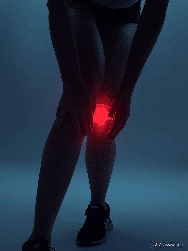
Runner's knee is one of the most common injuries among physically active individuals. As the name suggests, this condition occurs primarily in track-and-field athletes, but it is also frequently encountered among enthusiasts of cycling, skiing, football, and handball. It is most commonly associated with the gradual accumulation of the effects of intense lower-limb training. This pathology belongs to the group of overuse syndromes affecting the iliotibial band (ITB). The iliotibial band is not an isolated anatomical structure, but a specialized thickening of the fascia lata running between the hip and the knee. At its proximal insertion, fibers of the gluteal muscles (gluteus maximus and medius) merge with those of the tensor fasciae latae. Its distal portion inserts into the lateral epicondyle of the femur and Gerdy's tubercle located on the tibia. Equally frequent accompanying conditions include patellofemoral pain syndrome or chondromalacia patellae. Undoubtedly, sports participation is the primary context for the onset of runner's knee. However, physical exertion alone does not cause the condition. Runner's knee frequently develops as a result of running on uneven surfaces, inadequate or improper warm-up routines, inappropriate footwear, or sudden excessive training loads. Recurrent microtrauma or incompletely rehabilitated previous injuries also elevate the risk. The underlying biomechanical mechanism involves disrupted patellar tracking, which over time generates excessive friction and load within the patellofemoral joint and triggers synovial inflammation. Additional contributing factors include overstretched or inhibited quadriceps muscles, lower extremity structural malalignments (flat feet, abnormal tarsal statics, excessive foot pronation), patellofemoral rhythm disturbances, muscular imbalance (tight triceps surae combined with weakened quadriceps), an elevated Q-angle, and ankle joint dysfunctions. Furthermore, non-athletic factors can also contribute, notably prolonged sitting with crossed legs, hormonal fluctuations during puberty, and excessive body weight, which compromises lower extremity joint stability.
Patellofemoral pain syndrome in runners: Clinical presentation and symptomatic profile
Runner’s knee, clinically referred to as patellofemoral pain syndrome, represents a constellation of distinctive symptoms rooted in biomechanical and structural dysfunction. The hallmark feature is pronounced pain, typically localized to the anterolateral aspect of the knee joint, which exacerbates during activities such as walking on level ground, descending stairs, running, transitioning into kneeling or squatting positions, and upon palpation of the affected joint. This pain arises from abnormal friction of the iliotibial band due to disruption of its physiological movement pathway. Symptom intensity peaks at approximately 30 degrees of knee flexion, where frictional forces are maximized. Additionally, pain may radiate to the patellar region or beneath it, potentially indicating concurrent patellofemoral conflict. Beyond pain, the syndrome manifests as swelling, joint contour distortion, a sensation of pressure or fullness in the inferior patellar area, and knee instability stemming from ligamentous laxity and compromised stabilizing structures. Importantly, certain symptoms may reflect alternative pathologies—for instance, anterior knee pain could originate from popliteus muscle dysfunction, whereas suprapatellar discomfort may correlate with hip abductor weakness, indirectly affecting knee mechanics.
Therapeutic approaches for "runner’s knee": from acute phase to advanced rehabilitation
Management of iliotibial band syndrome (commonly referred to as "runner’s knee") is contingent upon the stage of pathology and necessitates a tailored, multidisciplinary approach. During the initial phase, pain modulation and anti-inflammatory strategies take precedence, with emphasis on safeguarding compromised soft tissues—including tendons, quadriceps musculature, and the patellar tendon apparatus. Therapeutic progression spans from immobilization and cryotherapy (Stage I) to graded loading and thermotherapy (Stage II), culminating in decisions regarding potential surgical intervention or activity modification (Stage III). Equally critical is primary and secondary prevention, encompassing biomechanical optimization, central stabilizer strengthening, and patient education on safe training protocols.
Structured rehabilitation exercise protocols for restoring full physical functionality
The rate of bodily recovery and the overall efficacy of applied therapeutic interventions are substantially contingent upon the consistent execution of tailored rehabilitation exercises. Of paramount importance is the patient’s active participation in the healing process—since greater dedication and perseverance in adhering to prescribed regimens correlate directly with more pronounced and enduring outcomes. During the preliminary phase, emphasis is placed on isometric exercises designed to elongate the quadriceps femoris muscle and the iliotibial band, supplemented by neuromuscular stimulation techniques. Subsequently, resistance-based training is incorporated to enhance joint stability and improve the flexibility of soft-tissue structures surrounding the compromised knee articulation. The next progression involves full lower-limb engagement exercises, which not only facilitate the restoration of muscular strength and dynamic capacity but also expand the restricted range of motion in the joint. The final stage entails functional training, wherein the affected limb is gradually subjected to full weight-bearing activities. Throughout the therapeutic continuum, exercise intensity is meticulously calibrated by a physiotherapy specialist in accordance with the patient’s individualized progress metrics.

mgr Tim Klein
✓Diets Premium Expert
Diets Premium team expert. Specializes in nutrition plans, strength sports, and training plans.
View Profile →